√70以上 dextrocardia ecg 213228-Dextrocardia ecg placement
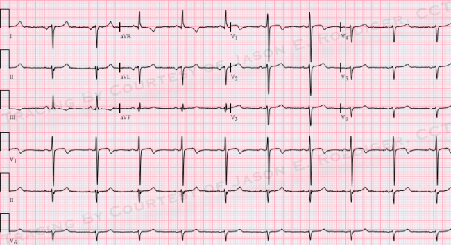
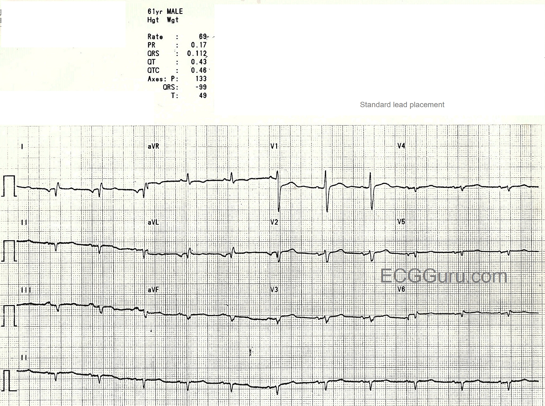
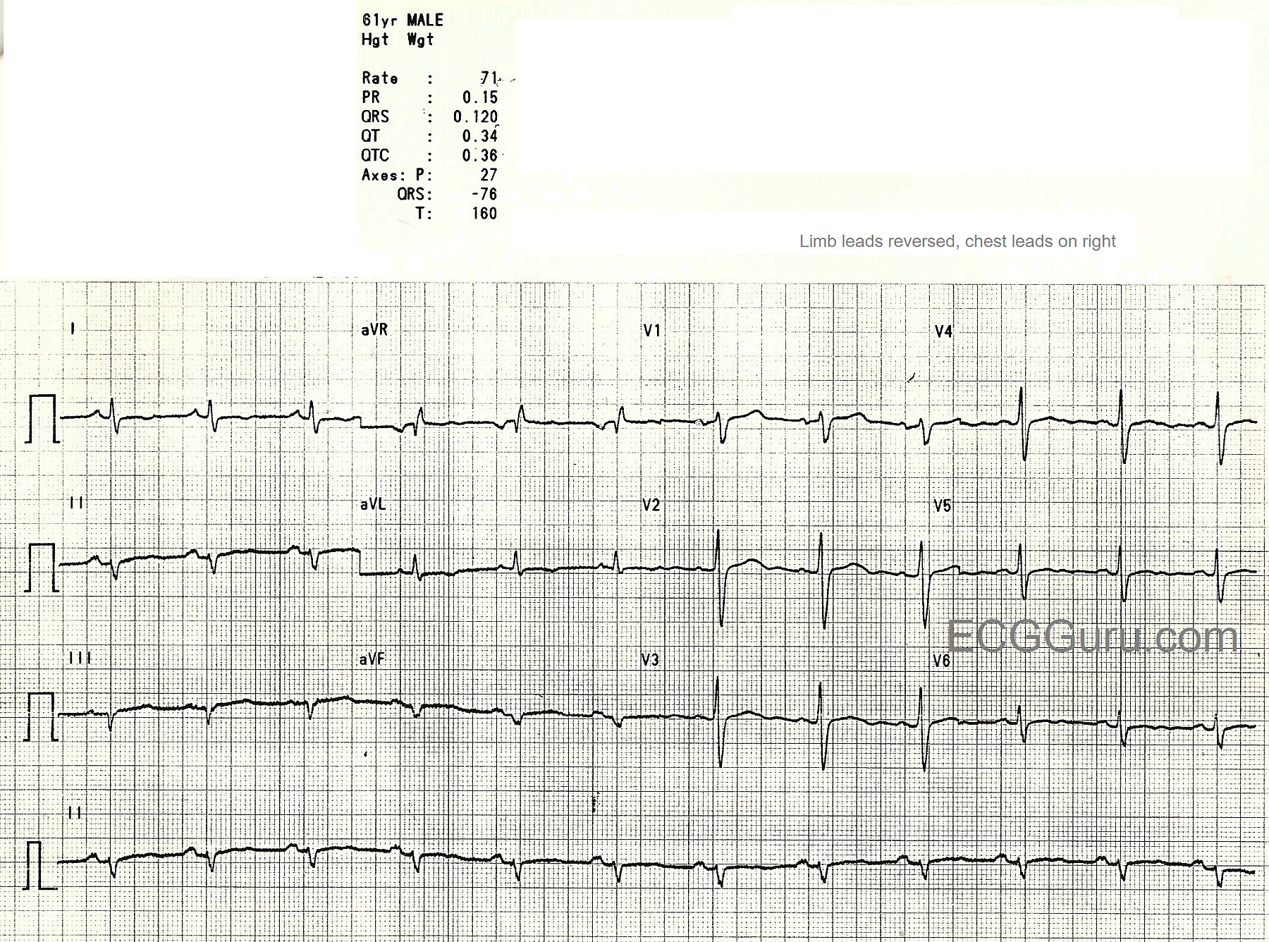
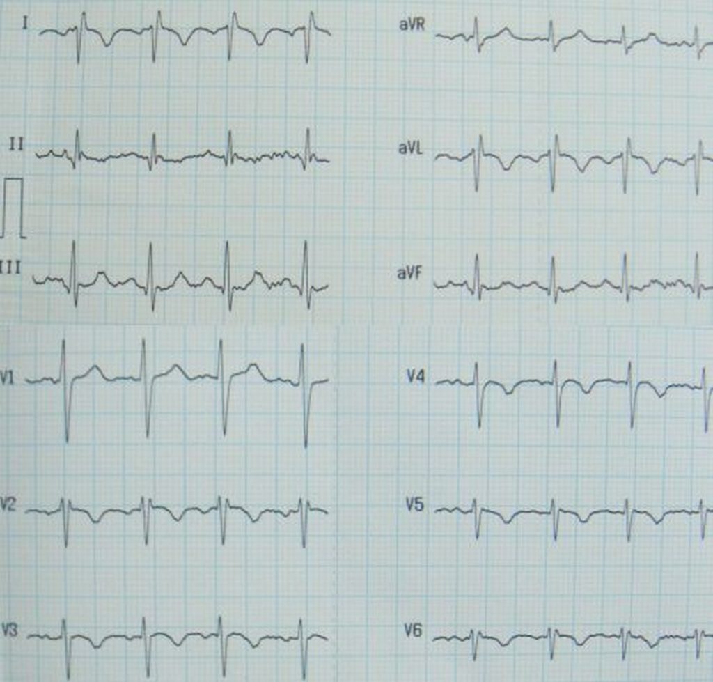
Dextrocardia This middleaged man presents to the Emergency Dept complaining of shortness of breath and fever Blood work, chest xray, and ECG are ordered When the ER Tech obtains the 12Lead ECG, he is puzzled by the following findingsDextrocardia is a condition in which the apex of the heart is directed towards the right side of the chest Dextrocardia can also cause the heart to develop in a mirror image of the normal heart In the more common types of dextrocardia, heart defects are present in addition to the abnormal location of the heartDextrocardia is defined as a cardiac position that is a mirror image of normal anatomy When the position of both the thoracic and abdominal viscera are reversed, this is referred to as dextrocardia with situs inversus (situs inversus totalis)
Dextrocardia Ecg Guru Instructor Resources
Dextrocardia ecg placement
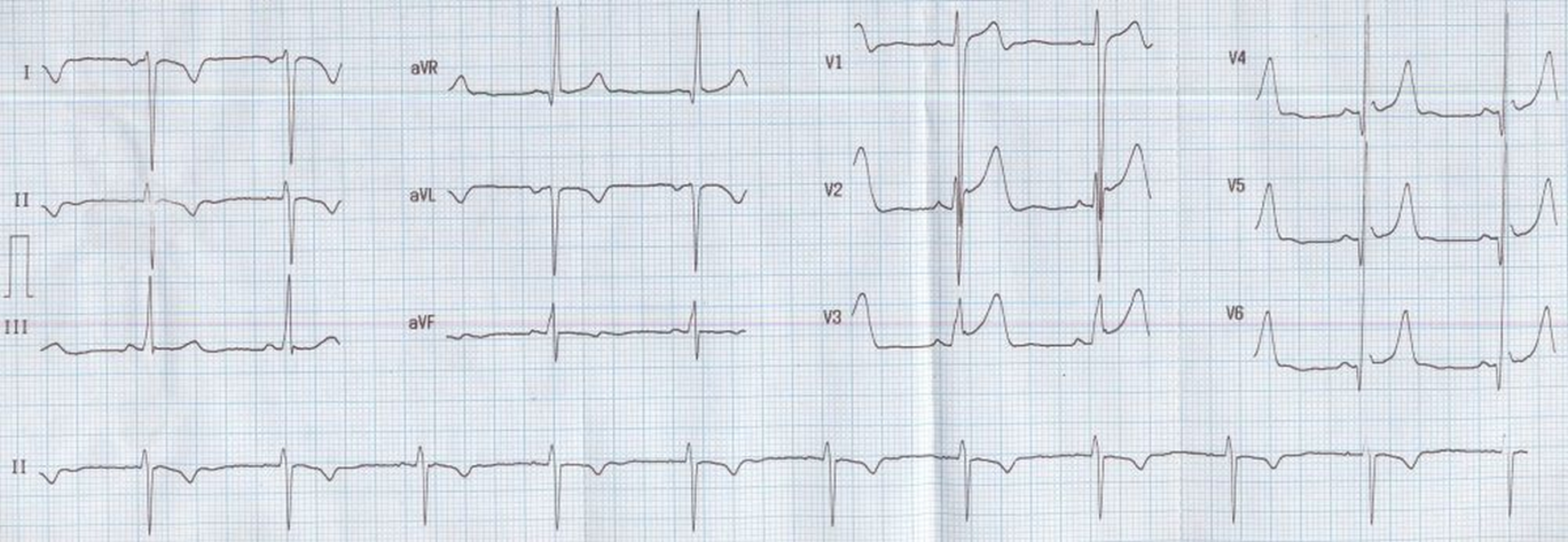
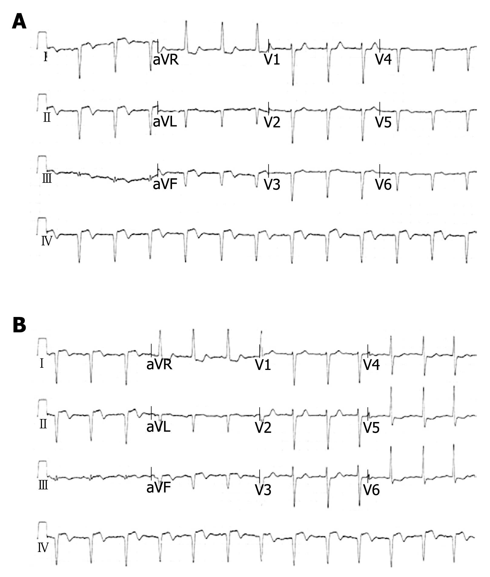
Dextrocardia ecg placement-ECG 4b With the diagnosis of dextrocardia, the chest electrodes were symmetrically placed on right hemithorax and the limb lead electrodes were reversed The new ECG now displays right bundle branch block and 1st degree AV block (above) P waves are normalized The wide S waves expected in lateral leads right during right bundle branch blockDextrocardia is a cardiac positional anomaly in which the heart is located in the right hemithorax with its basetoapex axis directed to the right and caudad The malposition is intrinsic to the heart and not caused by extracardiac abnormalities


Q Tbn And9gcrfdttvbw4q6dki2goyazrgsho3ust9cpk Cfyrmjrgsadjagej Usqp Cau
With dextrocardia situs inversus, the heart is a mirror image of its normal position dextrocardia situs totalis all visceral organs are mirrored So, if you see a ECG and it looks as if your leads are all reversed, consider that maybe the heart is Dextrocardia is a rare condition and occurs in approximately 1 in 12,000 peopleChest xrays and an ECG (echocardiogram) may be used to determine which type of dextrocardia is present Isolated dextrocardia (ie without any other associated heart defects) is a rare condition and occurs with equal frequency in males and femalesECG leads Dextrocardiacan usually be distinguished by noting that the precordial V leads do not have a normal R wave transition and by recording right sided chest leads which should be a mirror image of the normal left sided leads
Dextrocardia ECG Example 1 Intervention 15 ACC/AHA/SCAI Focused Update on Primary PCI for Patients With STEMIDextrocardia situs inversus refers to the heart is a condition where the heart is abnormally located on the right side of the thorax Dextrocardia situs inversus totalis is a condition where the heart is abnormally located on the right side of the thorax • ECG findings associated with dextrocardia includeECG Criteria & Findings By DX;
This Video Lecture Explains the ECG Changes in DextrocardiaLeads were then reversed and another ECG was taken as shown below With the precordial leads placed on the right side of the heart, the ECG now shows an ST elevation lateral infarction with reciprocal ST changes inferiorly It was an important pick as we have gone from ST depression to an ST elevation MI The dextrocardia was confirmed by chestECG Features of Dextrocardia Right axis deviation;


Q Tbn And9gcrfdttvbw4q6dki2goyazrgsho3ust9cpk Cfyrmjrgsadjagej Usqp Cau


Dextrocardia Dextroposition Ekg Ecg Ankara Kardiyoloji Kalp Hastaliklari Mete Alpaslan Doktorekg Com
OCCLUSION MI – STEMI & BEYOND;The Most Time Sensitive ECG Saves!ECG Dextrocardia 1 ECG OF THEECG OF THE WEEKWEEK DrBGowrishankarDrBGowrishankar ProfGSundaramurthy's unitProfGSundaramurthy's unit 2 Clinical HistoryClinical History 30 yrs old male30 yrs old male h/o cough, cold x 3 daysh/o cough, cold x 3 days Severe myalgia x 1 daySevere myalgia x 1 day O/EO/E AfebrileAfebrile Vitals


Abnormal P Wave And Qrs Complex Axes When Differential Diagnoses Overlap Annals Of Emergency Medicine


Q Tbn And9gctzscjoep Kco1qtfne4ylkbblnfvshztgttqdoa2oz4g Pcvu Usqp Cau
Dextrocardia is a rare condition where the heart points to the left side of the body instead of the right It does not show symptoms and is rarely lifethreatening all though it can occurMaster ECG interpretation from our nationallyknown educators Join Today!Dextrocardia may cause no symptoms if the heart is normal Usually, however the skin has a bluish color (cyanosis), there is difficulty breathing, fatigue, pale color, jaundice, recurrent sinus and lung infections and failure to grow What are dextrocardia care options?


Dr Smith S Ecg Blog A Woman With New Dyspnea Is The Extreme Left Axis Deviation With Negative T Wave In Lead Iii Suggestive Of Rv Strain


Myocardial Revascularization In Patient With Situs Inversus Totalis Case Report
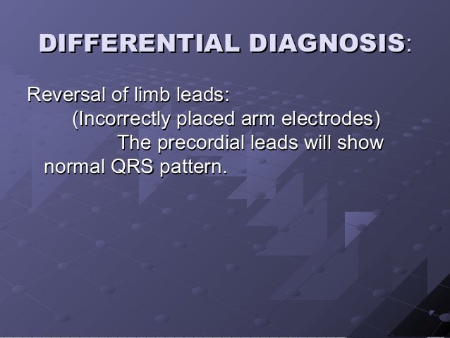
ECG study card for dextrocardia #Diagnosis #Cardiology #EKG #Dextrocardia #ECGEducator Contributed by Dr Gerald Diaz @GeraldMD Board Certified Internal Medicine Hospitalist, GrepMed Editor in Chief 🇵🇭 🇺🇸 Sign up for an account to like, bookmark and upload images to contribute to our community platformDextrocardia will show an R wave inversion, whereas lead reversal will not The bottom EKG shows marked right axis deviation and loss of voltage across the precordium There are also inverted P waves in leads I and aVL The differential for inverted P waves in lead I and aVL is Dextrocardia vs Reversed Arm LeadsOCCLUSION MI – STEMI & BEYOND;


A 21 Year Old College Student With Chest Distress And Ecg Mimicking Dextrocardia Circulation


Ecg Interpretation Ecg Blog 175 Lead Reversal Lateral Mi Dextrocardia
When dextrocardia occurs without situs inversus, when the visceral situs isinversus, when the visceral situs is indeterminate (indeterminate (situs ambigussitus ambigus) or if isolated) or if isolated levocardia is present complex multiplelevocardia is present complex multiple anomalies are usually presentanomalies are usually present 33Dextrocardia with situs inversus is a condition that is characterized by abnormal positioning of the heart and other internal organsIn people affected by dextrocardia, the tip of the heart points towards the right side of the chest instead of the left sideSitus inversus refers to the mirrorimage reversal of the organs in the chest and abdominal cavityFig 5B —Images from ECGgated CT scan of 52yearold woman with dextrocardia, situs inversus, and congenitally corrected transposition of great arteries (TGA) Axial image at level of cardiac chambers shows that morphologic left atrium (LA) is connected to a morphologic right ventricle (RV), distinguished by prominent trabeculations along


Dextrocardia Dextroposition Ekg Ecg Ankara Kardiyoloji Kalp Hastaliklari Mete Alpaslan Doktorekg Com


Electrocardiogram With Sinus Rhythm With Dextrocardia Nonspecific T Download Scientific Diagram
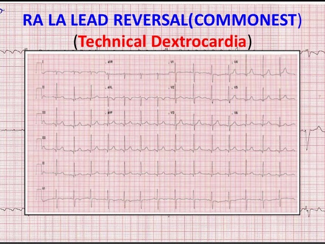
Dextrocardia with Situs Inversus is a rare heart condition characterized by abnormal positioning of the heart In this condition, the tip of the heart (apex) is positioned on the right side of the chest Additionally, the position of the heart chambers as well as the visceral organs such as the liver and spleen is reversed (situs inversus)When every tracing from an ECG machine exhibits this pattern it can be due to mislabeled ECG leads Dextrocardia can usually be distinguished by noting that the precordial V leads do not have a normal R wave transition and by recording right sided chest leads which should be a mirror image of the normal left sided leadsDextrocardia is a condition in which the heart is pointed toward the right side of the chest Normally, the heart points toward the left The condition is present at birth (congenital)


Dextrocardia Standard 12 Lead Ecg Good Heart Math Teaching


Dextrocardia Electrocardiogram Wikidoc
Dextrocardia is a rare condition in which the heart is located in the right side of the chest instead of the left Dextrocardia is usually present from birth (congenital) There are several types of dextrocardia The simplest type occurs when the shape and structure of the heart is a mirror image of a normal heartWith dextrocardia situs inversus, the heart is a mirror image of its normal position dextrocardia situs totalis all visceral organs are mirrored So, if you see a ECG and it looks as if your leads are all reversed, consider that maybe the heart isDextrocardia is a rare congenital heart condition that is characterized by presence of heart on right side instead of the normal left side It is estimated that less than 1% of the people may be born with Dextrocardia Quick facts about Dextrocardia The cause of Dextrocardia is not known Only right side location of the heart


Dextrocardia Ecg Guru Instructor Resources


Electrocardiography For Healthcare Professionals Ppt Download
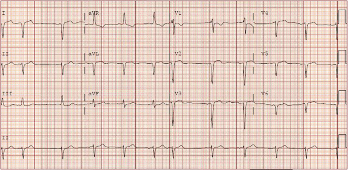
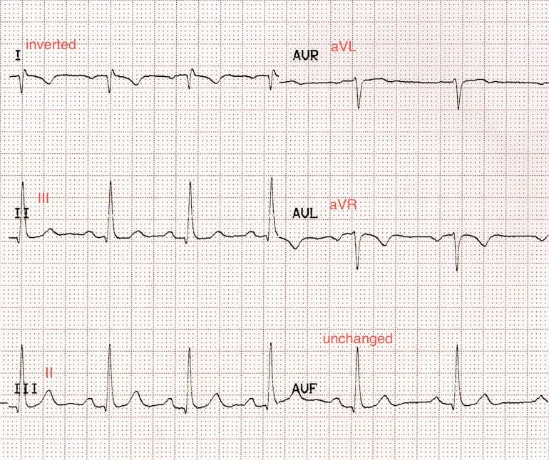
ECG The most prominent abnormality recognized on ECG is the "global inversion" of standard (limb) lead I, with inversion of the P wave, QRS complex, and T wave, suggestive of either dextrocardia or limb lead misplacement (the RA, or right arm, lead was placed on the left and vice versa) Use of the precordial leads allows discrimination ofPositive QRS complexes (with upright P and T waves) in aVR;RESPONSE TO ECG CHALLENGE The apex of the heart located at the right side of the chest is a reliable sign of dextrocardia


Electrocardiogram In Dextrocardia 25 Mm S 10 Mm Mv A Conventional Download Scientific Diagram


Dextrocardia Dextroposition Ekg Ecg Ankara Kardiyoloji Kalp Hastaliklari Mete Alpaslan Doktorekg Com
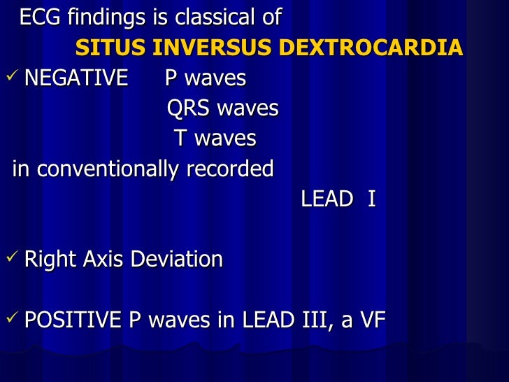
EKG Findings for Dextrocardia To recognize the EKG changes associated with a dextrocardia, it is important to have a clear understanding of the electrical axis • Global negativity in lead I (a negative Pwave, QRS complex and Twave) • Positively deflected QRS complex in aVRGet a full year access for only $26!ECG Diagnosis Dextrocardia ECG Diagnosis Dextrocardia ECG Diagnosis Dextrocardia Perm J 19; doi /TPP/144 Epub 19 Aug 15 Authors Cameron Mozayan 1 , Joel T Levis 1 2 3 Affiliations 1 Department of Emergency Medicine, Stanford University, CA 2 Department of


Dextrocardia Types Dextrocardia Situs Inversus Causes Symptoms Ecg Complications


Ecg Challenge 4 The Axis
Dextrocardia1 On a standard ECG with normal heart placement, lead I will almost always be positive and lead aVR negative with any supraventricular rhythm The finding of a positively deflected QRS complex in aVR and a negatively deflected ORS complex in lead I should cause concern The most common cause for this finding is reversedDextrocardia The heart is reversed and is in the right side of the chest rather than in its normal location on the left This is a true anatomic reversal With dextrocardia, for example, the apex (tip) of the heart points to the right rather than (as is normal) to the leftDextrocardia is a rare condition (around 1 in 12,000 births) that can be associated with a wider Heterotaxy Syndrome that can affect the heart, lungs, liver, spleen and intestines Children with dextrocardia require referral to multiple specialities and consideration for antibacterial prophylaxis if they are at risk of functional hyposplenia or


Dextrocardia Dextroposition Ekg Ecg Ankara Kardiyoloji Kalp Hastaliklari Mete Alpaslan Doktorekg Com


Ecg Primer For The Cath What Does A Tall R Wave In V1 Mean The Four Categories Approach Cath Lab Digest
The Most Time Sensitive ECG Saves!This Video Lecture Explains the ECG Changes in DextrocardiaDextrocardia with situs inversus is a condition that is characterized by abnormal positioning of the heart and other internal organs In people affected by dextrocardia, the tip of the heart points towards the right side of the chest instead of the left side


Dextrocardia Litfl Ecg Library Diagnosis


Ppt Ecg Etc Miscellaneous Ecgs Powerpoint Presentation Free Download Id
Dextrocardia is a rare heart condition in which your heart points toward the right side of your chest instead of the left side Dextrocardia is congenital, which means people are born with thisECG Criteria & Findings By DX;Lead I inversion of all complexes, aka 'global negativity' (inverted P wave, negative QRS, inverted T wave)


Dextrocardia With Cor Biloculare In A 44 Year Old Women


Case Report New Onset Heart Failure In Congenitally Corrected Transposition Of The Great Arteries Dextrocardia And Situs Inversus In An Octogenarian Journal Of Congenital Cardiology Full Text
Fig 5B —Images from ECGgated CT scan of 52yearold woman with dextrocardia, situs inversus, and congenitally corrected transposition of great arteries (TGA) Axial image at level of cardiac chambers shows that morphologic left atrium (LA) is connected to a morphologic right ventricle (RV), distinguished by prominent trabeculations alongDextrocardia is a congenital cardiac malposition in which the heart is situated on the right side of the body (dextroversion) with the cardiac apex pointing to the rightDextrocardia is a rare congenital heart condition that is characterized by presence of heart on right side instead of the normal left side It is estimated that less than 1% of the people may be born with Dextrocardia Quick facts about Dextrocardia The cause of Dextrocardia is not known


Wm19 Dextrocardia Flashcards Quizlet


Http Proceedings Med Ucla Edu Wp Content Uploads 17 01 Dextrocardia With Situs Pdf
• With vector manipulation ECG machine creates aVR, aVL, & aVF Hexaxial System • Used to determine electrical axis • What is the normal axis for the heart?Dextrocardia is a condition where the heart is located on the right side of the body, as opposed to the left This condition is typically the result of a birth defect and doesn't usually cause any problems Patients with Dextrocardia are often undiagnosed until they receive their first chest xray or ECGDocumenting the ECG results in the notes 1 Document the time and date that the ECG was performed as this may be significantly different from the time you are documenting 2 Write the indication for the ECG (eg chest pain, tachycardia) 3 Document your interpretation of the ECG (see our guide to interpreting an ECG) Rate;


Dextrocardia Guideline Paediatric Innovation Education And Research Network


Ekg Ecg Dextrocardia The Ekg Guy Www Ekg Md Youtube
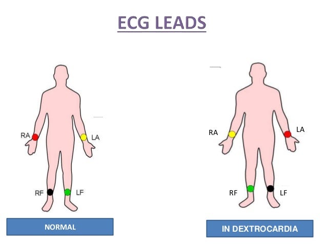
Dextrocardia The heart is reversed and is in the right side of the chest rather than in its normal location on the left This is a true anatomic reversal With dextrocardia, for example, the apex (tip) of the heart points to the right rather than (as is normal) to the leftECG leads must be placed in reversed positions on a person with dextrocardia In addition, when defibrillating someone with dextrocardia, the pads should be placed in reverse positions That is, instead of upper right and lower left, pads should be placed upper left and lower rightDextrocardia (from Latin dexter, meaning "right," and Greek kardia, meaning "heart") is a rare congenital condition in which the apex of the heart is located on the right side of the body There are two main types of dextrocardia dextrocardia of embryonic arrest (also known as isolated dextrocardia) citation needed and dextrocardia situs inversus


Left Bundle Branch Block Ecg 4 Learntheheart Com


Dextrocardia Can You Pick It On Ecg Resus
• 30 to 90 Electrical Axis Right Axis Deviation RVH Left posterior hemiblock Dextrocardia Ectopic ventricular beats and rhythms Left axis deviation Left Anterior


Living With Dextrocardia Living With Dextrocardia


12 Lead Ecg Lead Placement Diagrams Ems 12 Lead


Dextrocardia Ecg Guru Instructor Resources


Q Tbn And9gcs2otayjotplwej6re1ibmqzzq1fnnumhmj6ae27tyco8s6eq D Usqp Cau


R Wave Litfl Ecg Library Basics


Www Jacc Org Doi Pdf 10 1016 J Jaccas 06 009


42wqzjtrc5ia M


Ecg Study Card For Dextrocardia Diagnosis Cardiology Grepmed


Dextro Cardia A Rare Case


A Complete Guide About Dextrocardia Its Ecg Interpretation Blogs Tricog


Ekg Lead Placement Worksheet Printable Worksheets And Activities For Teachers Parents Tutors And Homeschool Families


Dextrocardia Dextroposition Ekg Ecg Ankara Kardiyoloji Kalp Hastaliklari Mete Alpaslan Doktorekg Com


Left Sided Ecg Of A 50 Year Old Man With Dextrocardia Situs Inversus Download Scientific Diagram
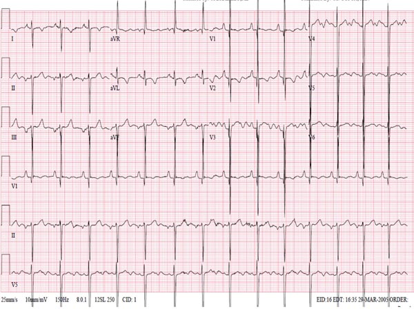


Cardiac Malpositions Including Heterotaxy Syndromes Thoracic Key


Figure 2 From Isolated Dextrocardia With Situs Solitus Dextroversion Semantic Scholar


Recognition Of Anterior Stemi In Dextrocardia And The Importance Of Right Sided Chest Leads Jacc Case Reports


Dextrocardia Or Leads Reversal Ecg


Dextrocardia And Proper Lead Placement Medic Madness


Ecg Rhythms Ectopic Atrial Rhythm In Dextrocardia


Dextrocardia Dextroposition Ekg Ecg Ankara Kardiyoloji Kalp Hastaliklari Mete Alpaslan Doktorekg Com


Incorrect Electrode Placement The Premier Ekg Resource For Medical Professionals Ekg Md Dr Anthony Kashou


Ecg Educator Blog Dextrocardia
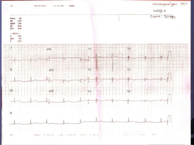


Ecg Dextrocardia
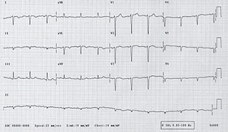


Percutaneous Coronary Intervention In Dextrocardia The British Journal Of Cardiology


Dextrocardia On Ekg See Corresponding Cxr Www Grepmed Com Images 4981 Grepmed
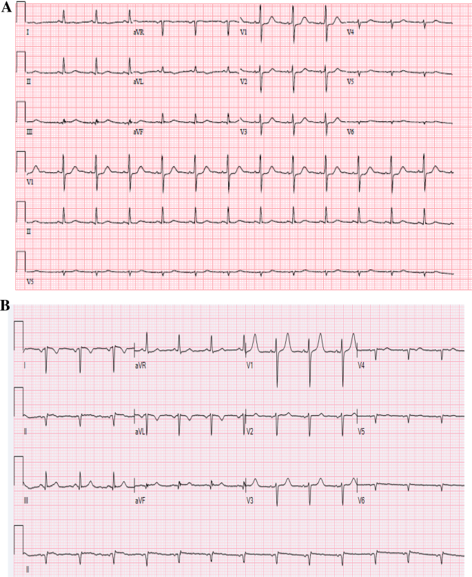


An Unusual Electrocardiogram Springerlink


Abnormal P Wave And Qrs Complex Axes When Differential Diagnoses Overlap Annals Of Emergency Medicine


Case Report New Onset Heart Failure In Congenitally Corrected Transposition Of The Great Arteries Dextrocardia And Situs Inversus In An Octogenarian Journal Of Congenital Cardiology Full Text


Dextrocardia Circulation


Lead Reversal Left Arm Right Arm Litfl Ecg Library Diagnosis


Ccu D E X T R O C A R D I A Dextrocardia Facebook


Arm Lead Inversion Technical Dextrocardia


Dextrocardia Ecg Guru Instructor Resources


Ecg Findings In Dextrocardia Cardiology Www Medicaltalk Net The Best Medical Forum For Medical Students And Doctors Worldwide


Dextrocardia On Ecg


Mirror Image Dextrocardia With Situs Inversus Circulation


Curso Ecg Dextrocardia


A Twist Of Fate Situs Inversus Totalis With Dextrocardia The American Journal Of Medicine


Ecg Class Keeping Ecgs Simple Dextrocardia Ecgclass Case 30


Inferior Myocardial Infarction In A Patient With Mirror Image Dextrocardia And Situs Inversus Totalis Sciencedirect


View Image


How Standard Transesophageal Echocardiography Views Change With Dextrocardia Raut Ms Maheshwari A Shad S Rachna G Ann Card Anaesth


Dextrocardia Dr Uri Ben Zur Asymptomatic Male Ekg Ecg Electrocardiogram Youtube


Ecg Cases 15 Tall R Wave In V1 Emergency Medicine Cases


The Role Of Electrocardiogram In The Diagnosis Of Dextrocardia With Mirror Image Atrial Arrangement And Ventricular Position In A Young Adult Nigerian In Ile Ife A Case Report Semantic Scholar




Ecg Artifacts And Pitfalls


Q Tbn And9gctpydl0 Xkxoiabceqks4h3g2d Mhvy1gate0cuqp3w2b Cs8po Usqp Cau


12 Lead Ecg Lead Placement Diagrams Ems 12 Lead


17 Ecg Of Dextrocardia Youtube


Kartagener Syndrome Dextrocardia Situs Inversus Immotile Cilia


Ecg Response Can You Make The Correct Morphology Pathology And Rhythm Diagnoses Circulation


Essentials Of Electrocardiography Ecg


Percutaneous Coronary Intervention For Acute Myocardial Infarction In A Patient With Dextrocardia


Learntheheart Com Dextrocardia Ecg Heart Located In Right Chest Negative P Qrs T In Lead I Low Voltage V3 V6 Usmle Http T Co Ssrk1hvr98


Dextrocardia On Ekg Limb Leads Look Like La Grepmed


Ecg Cardiac Malpositions


Myocardial Revascularization In A Patient With Situs Inversus Totalis


Dextrocardia Electrocardiogram Wikidoc


Www Jacc Org Doi Pdf 10 1016 J Jaccas 06 009


Dextrocardia Can You Pick It On Ecg Resus


Dextrocardia Wikipedia


Dextrocardia And Acute Anterior Myocardial Infarction Cardiology Www Medicaltalk Net The Best Medical Forum For Medical Students And Doctors Worldwide


Http Proceedings Med Ucla Edu Wp Content Uploads 17 01 Dextrocardia With Situs Pdf


Electrocardiographic Findings In A Woman With Dextrocardia And Cyanosis Jama Internal Medicine X Mol


Ecg Features Of Dextrocardia Right Medical Infopedia By Dr Ali Shahzad Malik Facebook


Ekg สำหร บ คนห วใจกล บด าน Dextrocardia ม ลน ธ เพอร เฟคไลฟ Facebook


Left Sided Ecg Of A 50 Year Old Man With Dextrocardia Situs Inversus Download Scientific Diagram


Ecg Dextrocardia


Electrocardiography Wikipedia


Bozwell Co Uk
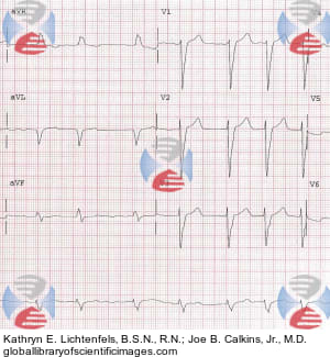


Dextrocardia Left Bundle Branch Block And Atrial Fibrillation Global Library Of Scientific Images


Gale Onefile Health And Medicine Document Electrocardiogram Read By The Computer As Arm Lead Reversal


コメント
コメントを投稿